I. Define the terms: organ, organ system and organism.
II. Name the eleven organ systems of the human body, identify the major organs, and give a major function of each system.
III. Define and demonstrate the anatomical position.
IV. Locate the anterior (ventral) and posterior (dorsal) surfaces for the body, hands, and feet.
V. Define the directional terms used in human anatomy.
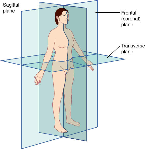
VI. Define sagittal, frontal, and transverse planes and distinguish between midsagittal (median) and parasagittal planes.
VII. Specify and describe the limits of the body cavities.
VIII. Describe how the abdominopelvic region is divided into either nine regions or four quadrants.
An organ is an anatomically distinct structure of the body composed of two or more tissue types. Each organ performs one or more specific physiological functions. An organ system is a group of organs that work together to perform major functions or meet physiological needs of the body.
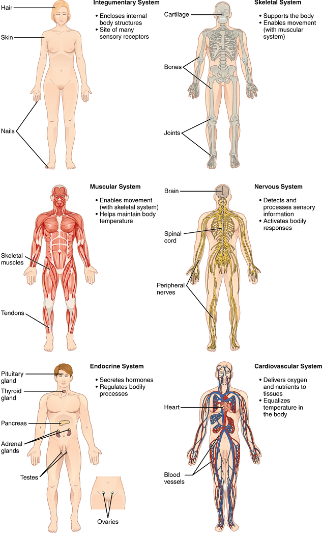
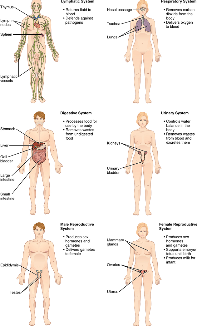
The human body contains eleven distinct organ systems (Figure 1.1 and Figure 1.2). Assigning organs to organ systems can be imprecise, since organs that “belong” to one system can also have functions integral to another system. In fact, most organs contribute to more than one system.
The organism level is the highest level of anatomical organization. An organism is a living being that has a cellular structure and can independently perform all physiologic functions necessary for life. In multicellular organisms, including humans, all cells, tissues, organs, and organ systems of the body work together to maintain the life and health of the organism.
Test Your Knowledge
Define the terms organ, organ system, and organism.
Anatomists and health care providers use terminology that can be bewildering to the uninitiated. However, the purpose of this language is not to confuse but rather to increase precision and reduce medical errors. For example, is a scar “above the wrist” located on the forearm two or three inches away from the hand? Or is it at the base of the hand? Is it on the palm side or back side? By using precise anatomical terminology, we eliminate ambiguity. Anatomical terms derive from Ancient Greek and Latin words. Because these languages are no longer used in everyday conversation, the meaning of their words does not change.
Anatomical terms are made up of roots, prefixes, and suffixes (Appendix II). The root of a term often refers to an organ, tissue, or condition, whereas the prefix or suffix often describes the root. For example, in the disorder hypertension, the prefix “hyper-” means “high” or “over,” and the root word “tension” refers to pressure, so the word “hypertension” refers to abnormally high blood pressure.
Test Your Knowledge
To further increase precision, anatomists standardize the way in which they view the body. Just as maps are normally oriented with north at the top, the standard body “map,” or anatomical position, is that of the body standing upright, with the feet parallel, shoulder width apart, and with toes forward. The upper limbs are held out to each side, and the palms of the hands face forward (Figure 1.3). Using this standard position reduces confusion. It does not matter how the body being described is oriented, the terms are used as if it is in anatomical position. For example, a scar in the “anterior (front) carpal (wrist) region” would be present on the palm side of the wrist. The term “anterior” would be used even if the hand were palm down on a table.
A body that is lying down is described as either prone or supine. Prone describes a face-down orientation, and supine describes a face-up orientation. These terms are sometimes used in describing the position of the body during specific physical examinations or surgical procedures.
Test Your Knowledge
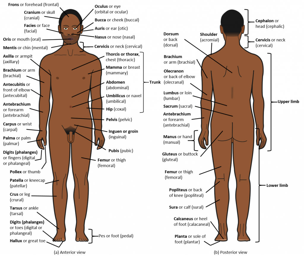
The human body’s numerous regions have specific terms to help increase precision (Figure 1.3). Notice that the term “brachium” or “arm” is reserved for the “upper arm,” and “antebrachium” or “forearm” is used rather than “lower arm.” Similarly, “femur” or “thigh” is correct, and “leg” or “crus” is reserved for the portion of the lower limb between the knee and the ankle. You will be able to describe the body’s regions using the terms from the figure.
Test Your Knowledge
Certain directional anatomical terms appear throughout all anatomy textbooks (Figure 1.4). These terms are essential for describing the relative locations of different body structures. For instance, an anatomist might describe one band of tissue as “inferior to” another, or a physician might describe a tumor as “superficial to” a deeper body structure. Commit these terms to memory to avoid confusion when you are studying or describing the locations of particular body parts.
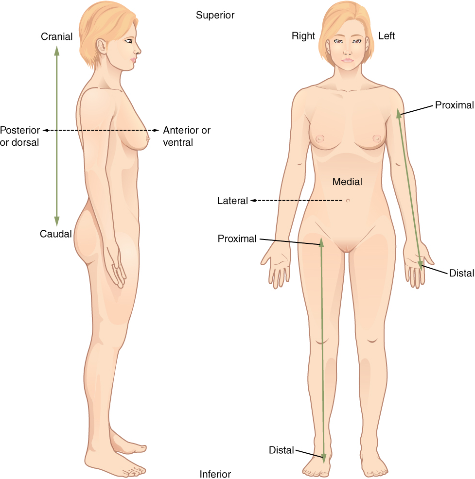
Deep describes a position farther from the surface of the body. The brain is deep to the skull.
Test Your Knowledge
1. Define each of the following terms and provide one complete sentence that correctly uses each term to describe the relative position of two or three body structures (as appropriate).
2. Distinguish between the terms prone and supine.
Sectioning, or cutting, is frequently used in the study of Anatomy. The body can be sectioned in various ways to produce a plane, this is a two-dimensional surface of a three-dimensional structure that has been cut. A body structure is often cut into thin sections before macroscopic viewing to allow visualization of the structure’s interior and assist with identification of local disease or infiltration, as these pathologies may not be obvious when observing the surface anatomy alone. Modern medical imaging devices enable clinicians to obtain “virtual sections” of living bodies. We call these scans. Body sections and scans can be correctly interpreted, however, only if the viewer understands the plane along which the section was made. A plane is an imaginary two-dimensional surface that passes through the body. There are three planes commonly referred to in anatomy and medicine (Figure 1.5).
Test Your Knowledge
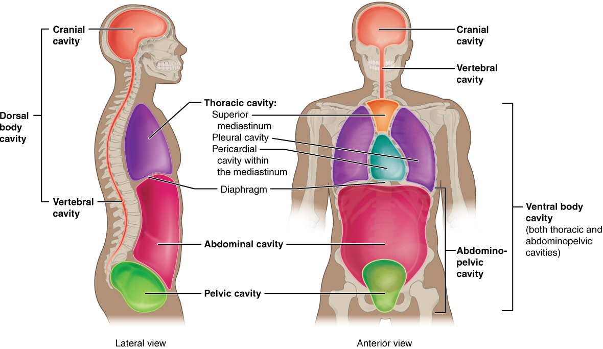
The body maintains its internal organization by means of membranes, sheaths, and other structures that separate compartments. The dorsal (posterior) cavity and the ventral (anterior) cavity are the largest body compartments (Figure 1.6). These cavities contain and protect delicate internal organs, and the ventral cavity allows for significant changes in the size and shape of the organs as they perform their functions. The lungs, heart, stomach, and intestines, for example, can expand and contract without distorting other tissues or disrupting the activity of nearby organs.
Subdivisions of the Posterior (Dorsal) and Anterior (Ventral) Cavities: The posterior (dorsal) and anterior (ventral) cavities are each subdivided into smaller cavities. In the posterior (dorsal) cavity, the cranial cavity houses the brain, and the spinal cavity (or vertebral cavity) encloses the spinal cord. Just as the brain and spinal cord make up a continuous, uninterrupted structure, the cranial and spinal cavities that house them are also continuous. The brain and spinal cord are protected by the bones of the skull and vertebral column and by cerebrospinal fluid, a colorless fluid produced by the brain, which cushions the brain and spinal cord within the posterior (dorsal) cavity.
The anterior (ventral) cavity has two main subdivisions: the thoracic cavity and the abdominopelvic cavity (Figure 1.6). The thoracic cavity is the more superior subdivision of the anterior cavity, and it is enclosed by the rib cage. The thoracic cavity contains the lungs and the heart, which is located in the mediastinum. The diaphragm forms the floor of the thoracic cavity and separates it from the more inferior abdominopelvic cavity. The abdominopelvic cavity is the largest cavity in the body. Although no membrane physically divides the abdominopelvic cavity, it can be useful to distinguish between the abdominal cavity, the division that houses the digestive organs, and the pelvic cavity, the division that houses the organs of reproduction.
Test Your Knowledge
Abdominopelvic Regions and Quadrants: To promote clear communication, for instance, about the location of a patient’s abdominal pain or a suspicious mass, health care providers typically divide up the abdominopelvic cavity into either nine regions or four quadrants (Figure 1.7).
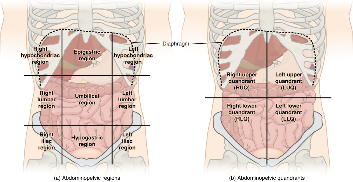
The simpler quadrants approach, which is also commonly used in medicine, subdivides the cavity with one horizontal and one vertical line that intersect at the patient’s umbilicus (navel). The quadrants approach are described as:
The more detailed regional approach subdivides the cavity with one horizontal line immediately inferior to the ribs and one immediately superior to the pelvis and two vertical lines drawn as if dropped from the midpoint of each clavicle (collarbone). There are nine resulting regions. These regions can be used to identify the location of abdominal organs more precisely. For example:
Membranes of the Anterior (Ventral) Body Cavity: A serous membrane (also referred to as a serosa) is one of the thin membranes that cover the walls and organs in the thoracic and abdominopelvic cavities. The parietal layers of the membranes line the walls of the body cavity (pariet- refers to a cavity wall). The visceral layer of the membrane covers the organs (the viscera). Between the parietal and visceral layers is a very thin, fluid-filled serous space, or cavity (Figure 1.8).
There are three serous cavities and their associated membranes. The pleura is the serous membrane that surrounds the lungs in the pleural cavity; the pericardium is the serous membrane that surrounds the heart in the pericardial cavity; and the peritoneum is the serous membrane that surrounds several organs in the abdominopelvic cavity.
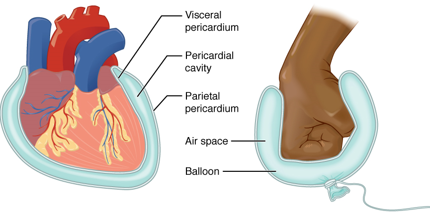
The serous membranes form fluid-filled sacs, or cavities, that cushion and reduce friction on internal organs when they move, such as when the lungs inflate or the heart beats. Both the parietal and visceral serosa secrete the thin, slippery serous fluid located within the serous cavities.
The pleural cavity reduces friction between the lungs and the body wall. Likewise, the pericardial cavity reduces friction between the heart and the wall of the pericardium. The peritoneal cavity reduces friction between the abdominal and pelvic organs and the body wall. Therefore, serous membranes provide additional protection to the viscera they enclose by reducing friction that could lead to inflammation of the organs.
Test Your Knowledge
1. Specify what organs are found in the following quadrants
2. Specify the location(s), within the nine abdominopelvic regions, of each of the following organs:
For the following questions, click the correct answer choice.
Figure 1.1: The image includes 6 systems of the body. The Integumentary System encloses internal body structures and the site of many sensory receptors. Consists of hair, skin, and nails. The Skeletal System supports the body and enables movement along with the muscular system. Consists of cartilage, bones, and joints. The Muscular System enables movement along with the skeletal system and helps maintain body temperature. Consists of skeletal muscles and tendons. The Nervous System detects and processes sensory information and activates bodily responses. Consists of brain, spinal cord, and peripheral nerves. The Endocrine System secretes hormones and regulates body processes. Consists of pituitary gland, thyroid gland, pancreas, adrenal gland, testes, and ovaries. The Cardiovascular System delivers oxygen and nutrients to tissues and equalizes temperature in the body. Consists of heart and blood vessels. [Return to image]
Figure 1.2: The image includes 6 more systems of the body. The Lymphatic System returns fluid to blood and defends against pathogens. Consists of thymus, lymph nodes, spleen, and lymphatic vessels. The Respiratory System removes carbon dioxide from the body and delivers oxygen to the blood. Consists of nasal passage, trachea, and lungs. The Digestive System processes food for use by the body and removes wastes from undigested food. Consists of stomach, liver, gallbladder, large intestine, and small intestine. The Urinary System controls water balance in the body and removes waste from the blood and secretes them. Consists of kidneys and urinary bladder. The Male Reproductive System produces sex hormones and gametes and delivers gametes to female. Consists of epididymis and testes. The Female Reproductive System produces sex hormones and gametes, supports the embryo/fetus until birth, and produces milk for the infant. Consists of mammary gland, ovaries, and uterus. [Return to image]
Figure 1.3: There are scientific terms for the body parts. The front of the body terms: forehead-frontal, skill-cranial, face-facial, eye-orbital, cheek-buccal, ear-otic, nose-nasal, neck-cervical, chest-thoracic, breast-mammary, mouth-oral, chin-mental, armpit-axillary, arm-brachial, elbow-antecubital, forearm-antebrachial, abdomen-abdominal, navel-umbilical, hip-coxal, wrist-carpal, thumb-pollex, palm-palmar, fingers-phalanges, kneecap-patellar, leg-crural, ankle-tarsal, great toe-hallux, foot-pedal, thigh-femoral, pubic-pubis, groin-inguinal, pelvic-pelvis, and hip-coxal. The back of the body terms: head-cephalic, neck-cervical, shoulder-acromial, back-dorsal, back of elbow-olecranal, loin-lumbar, sacrum-sacral, hand-manual, buttock-gluteal, back of knee-popliteal, calf-sural, and heel of foot-calacaneal. [Return to image]
Figure 1.4 This illustration shows an anterior and posterior view of the human body. The cranial region encompasses the upper part of the head, while the facial region encompasses the lower half of the head beginning below the ears. The eyes are referred to as the ocular region. The cheeks are referred to as the buccal region. The ears are referred to as the auricle or otic region. The nose is referred to as the nasal region. The chin is referred to as the mental region. The neck is referred to as the cervical region. The trunk of the body contains, from superior to inferior, the thoracic region encompassing the chest, the mammary region encompassing each breast, the abdominal region encompassing the stomach area, the coxal region encompassing the beltline, and the pubic region encompassing the area above the genitals. The umbilicus, or naval, is located at the center of the abdomen. The pelvis and legs contain, from superior to inferior, the inguinal or groin region between the legs and the genitals, the pubic region surrounding the genitals, the femoral region encompassing the thighs, the patellar region encompassing the knee, the crural region encompassing the lower leg, the tarsal region encompassing the ankle, the pedal region encompassing the foot, and the digital/phalangeal region encompassing the toes. The great toe is referred to as the hallux. The regions of the upper limbs, from superior to inferior, are the axillary region encompassing the armpit, the brachial region encompassing the upper arm, the antecubital region encompassing the front of the elbow, the antebrachial region encompassing the forearm, the carpal region encompassing the wrist, the palmar region encompassing the palm, and the digital/phalangeal region encompassing the fingers. The thumb is referred to as the pollux. The posterior view contains, from superior to inferior, the cervical region encompassing the neck, the dorsal region encompassing the upper back, and the lumbar region encompassing the lower back. The regions of the back of the arms, from superior to inferior, include the cervical region encompassing the neck, acromial region encompassing the shoulder, the brachial region encompassing the upper arm, the olecranal region encompassing the back of the elbow, the antebrachial region encompassing the back of the arm, and the manual region encompassing the palm of the hand. The posterior regions of the legs, from superior to inferior, include the gluteal region encompassing the buttocks, the femoral region encompassing the thigh, the popliteus region encompassing the back of the knee, the sural region encompassing the back of the lower leg, and the plantar region encompassing the sole of the foot. Some regions are combined into larger regions. These include the trunk, which is a combination of the thoracic, mammary, abdominal, naval, and coxal regions. The cephalic region is a combination of all of the head regions. The upper limb region is a combination of all of the arm regions. The lower limb region is a combination of all of the leg regions. [Return to image.]
Figure 1.5 The body can be divided into three planes. Coronal (frontal) plane: separates the front (anterior) and back (posterior) of the body. Sagittal (longitudinal) plane: separates the left and right sides of the body. Transverse (axial) plane: separates the upper (superior) and lower (inferior) halves of the body. [Return to image.]
Figure 1.6 Medical and crime shows have made body cavities all too familiar, and anatomically speaking, these spaces are very important, providing housing and protection for vital organs. The following list identifies the cavities and subcavities of the human body: